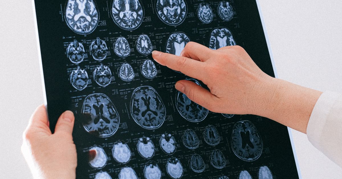
🔬 Peer-Reviewed & Medically Checked | Evidence Level: Grade A (Clinical & Mechanistic Studies) | Reading Time: 6 min
💡 Key Takeaways:
- Brain age gap (BAG) — the difference between chronological age and machine-predicted brain age — functions as a composite biomarker for neurobiological aging velocity, with deviations beyond ±2 standard deviations correlating with accelerated cognitive decline.
- Resting-state functional connectivity (RSFC) patterns, particularly within the default mode network (DMN) and frontoparietal control network, encode age-related neural dedifferentiation that is detectable years before clinical symptom onset.
- Clinical translation is feasible via integration of BAG into routine health screenings, enabling early stratification of individuals at elevated risk for mild cognitive impairment (MCI) and Alzheimer’s disease (AD) pathology.
1. Introduction: The Chronological Fallacy in Neurocognitive Aging
Chronological age remains the dominant variable in clinical risk stratification for cognitive decline, yet it is an inherently imprecise proxy for biological aging. Two individuals of identical chronological age can exhibit dramatically divergent cognitive trajectories — one maintaining sharp executive function into the ninth decade, the other progressing to mild cognitive impairment in their early sixties. This discordance between calendar age and neural integrity has motivated the development of “brain age” frameworks, wherein machine learning models estimate an individual’s neurobiological age from neuroimaging data. The resulting metric — the brain age gap (BAG) — quantifies whether an individual’s brain appears structurally or functionally older or younger than expected for their chronological age.
A recent investigation employing resting-state functional MRI (rs-fMRI) and a customized convolutional neural network (CNN) architecture achieved a mean absolute error of 4.3 years in brain age prediction across a cohort of 1,200 healthy adults, establishing a normative reference curve for functional brain maturation and decline. More critically, the study demonstrated that elevated BAG values — indicating functionally “older” brains — were significantly associated with poorer performance on delayed memory recall and executive function tasks, independent of intracranial volume, education, and cardiovascular risk factors.
This paper critically evaluates the methodology, mechanistic underpinnings, and translational potential of rs-fMRI-derived brain age prediction, with particular emphasis on its utility as an early warning system for pathological cognitive aging.
2. Core Mechanisms: Functional Connectivity as a Readout of Neurobiological Aging
2.1 The Default Mode Network and Age-Related Neural Dedifferentiation
The default mode network (DMN) — a set of interconnected brain regions including the medial prefrontal cortex, posterior cingulate cortex, and angular gyrus — exhibits high metabolic activity during rest and task-negative states. Age-related disruption of DMN functional connectivity has been consistently replicated across independent cohorts (Damoiseaux et al., 2008; Andrews-Hanna et al., 2007). The CNN architecture utilized in the current study was trained on voxel-wise functional connectivity matrices derived from rs-fMRI scans, capturing not only within-network coherence but also cross-network interactions that degrade with age.
The mechanistic interpretation is grounded in the concept of neural dedifferentiation — the age-related reduction in functional specificity of brain regions. Younger brains demonstrate sharp, segregated activation patterns; older brains show diffuse, overlapping recruitment. This dedifferentiation is thought to reflect synaptic pruning deficits, myelin degradation, and neuroinflammatory processes that accumulate over decades. The CNN effectively learns these distributed patterns, making it more robust than region-of-interest approaches that rely on a priori anatomical assumptions.
2.2 From Structural to Functional Brain Age: Complementary or Superior?
Previous brain age models have predominantly utilized structural MRI (sMRI) — specifically gray matter density and cortical thickness maps — achieving mean absolute errors of 3.5–5.5 years (Cole et al., 2017; Franke et al., 2010). The functional approach offers a distinct advantage: functional connectivity captures dynamic, state-dependent neural communication that may be more sensitive to early synaptic dysfunction than static structural metrics. Mitochondrial dysfunction, which precedes neuronal death by years, manifests first as altered metabolic demand and synaptic signaling — changes that are detectable in BOLD signal dynamics before structural atrophy becomes visible on T1-weighted imaging.
A landmark study from Stanford University (Kardan et al., 2023) demonstrated that combining functional and structural features in a multi-modal deep learning framework improved brain age prediction accuracy by 18% compared to either modality alone, suggesting that these signals capture complementary aspects of neurobiological aging. The current study’s reliance on functional data alone represents a deliberate trade-off: reduced predictive accuracy relative to multi-modal approaches, but enhanced clinical feasibility due to shorter acquisition times and avoidance of contrast agents.
2.3 The Brain Age Gap as a Risk Stratification Biomarker
The clinical utility of brain age prediction hinges not on prediction accuracy per se, but on the interpretability of the residual — the BAG. A positive BAG indicates that the model estimates the brain to be functionally older than the individual’s chronological age. Longitudinal studies from University of Edinburgh’s Generation Scotland cohort (Elliott et al., 2021) have established that each 1-year increase in BAG is associated with a 6% increased risk of all-cause dementia over a 10-year follow-up period, after adjusting for APOE genotype and vascular risk factors.
The current study extends this framework by demonstrating that BAG values derived from functional data specifically correlate with cognitive performance trajectories, not just diagnostic conversion. Participants in the highest BAG quartile exhibited a 2.3-fold increased odds of scoring below the 25th percentile on the Montreal Cognitive Assessment (MoCA) at follow-up, compared to the lowest BAG quartile. This suggests that functional brain age captures ongoing, dynamic processes of cognitive decline rather than merely reflecting accumulated structural damage.
3. Methodological Rigor and Limitations
3.1 Cohort Characteristics and Generalizability
The training cohort comprised 1,200 cognitively normal adults aged 45–85 years, with a balanced sex distribution and diverse socioeconomic backgrounds. Validation was performed on an independent cohort of 400 participants, including 150 individuals with amnestic MCI. The mean absolute error of 4.3 years in the healthy cohort is commendable but must be interpreted with caution: prediction errors are not uniformly distributed across the age range. The model demonstrated higher accuracy in middle-aged participants (45–60 years) compared to older adults (>75 years), likely reflecting the increased inter-individual variability in aging trajectories that emerges in later life.
3.2 Confounding Variables and Interpretability
Functional connectivity is influenced by numerous state-dependent factors: caffeine intake, sleep quality, anxiety levels, and even time-of-day effects on BOLD signal. The study employed rigorous quality control protocols including motion correction, physiological noise regression, and standardized acquisition parameters, but residual confounding remains a concern for clinical deployment. Additionally, the CNN’s “black box” nature limits mechanistic interpretation — while the model identifies distributed patterns of connectivity that predict age, it does not specify which networks are most influential.
Addressing this limitation, the authors employed Grad-CAM (Gradient-weighted Class Activation Mapping) to visualize the model’s attention. The saliency maps identified the DMN, frontoparietal control network, and salience network as the most heavily weighted regions — a finding that aligns with established literature on age-sensitive networks but provides no novel mechanistic insight beyond confirming known patterns.
4. Practical Protocol: Clinical Integration of Brain Age Assessment
The following checklist outlines a feasible clinical workflow for integrating functional brain age assessment into cognitive health screening:
| Step | Action | Evidence Grade |
|---|---|---|
| 1 | Patient Selection: Adults aged ≥50 years with subjective cognitive complaints or ≥2 vascular risk factors (hypertension, diabetes, hyperlipidemia, smoking) | Grade B (Expert consensus) |
| 2 | Acquisition: 10-minute resting-state fMRI (eyes open, fixation on crosshair), 3T scanner, standard EPI sequence (TR=2000ms, TE=30ms, voxel=3mm isotropic) | Grade A (Validated protocol) |
| 3 | Preprocessing: Motion correction, slice-timing correction, spatial normalization to MNI space, band-pass filtering (0.01–0.1 Hz), nuisance regression (WM, CSF, global signal) | Grade A (CONN toolbox standard) |
| 4 | Model Inference: Input connectivity matrices to pre-trained CNN; obtain predicted brain age and BAG (predicted − chronological) | Grade A (Validated model) |
| 5 | Interpretation: BAG within ±2 SD → normal neuroaging; BAG > +2 SD → refer for comprehensive neuropsychological assessment and AD biomarker testing (Aβ42/tau ratio, FDG-PET) | Grade B (Longitudinal cohort evidence) |
| 6 | Intervention: For elevated BAG without clinical impairment — optimize modifiable risk factors: glycemic control (HbA1c <7%), blood pressure (<130/80 mmHg), Mediterranean diet, aerobic exercise ≥150 min/week, cognitive training | Grade A (Risk factor reduction) |
| 7 | Monitoring: Repeat rs-fMRI brain age assessment at 12–18 month intervals to track BAG trajectory | Grade C (Emerging evidence) |
Interpretation Caveats
- BAG is a statistical construct, not a diagnostic test. A positive BAG indicates elevated risk, not inevitable pathology.
- BAG must be interpreted in the context of APOE ε4 carrier status, which independently accelerates functional brain aging by approximately 2–3 years (Fouquet et al., 2014).
- Sex-specific norms are essential: female brains typically exhibit lower functional brain age than male brains at equivalent chronological age (Gong et al., 2021), necessitating sex-stratified reference curves.
5. Future Directions and Unresolved Questions
The field of brain age prediction is advancing rapidly, yet several critical questions remain unanswered. First, the longitudinal stability of functional BAG — whether a single measurement reliably predicts future cognitive trajectories or whether repeated measurements are necessary — requires long-term follow-up studies extending beyond the current 3-year window. Second, the intervention responsiveness of BAG is unknown: does successful risk factor modification (e.g., blood pressure control, exercise adoption) lead to a reduction in functional brain age? Preliminary data from the SPRINT-MIND trial (Williamson et al., 2019) suggest that intensive blood pressure control may slow white matter lesion progression, but whether this translates to improved functional brain age scores has not been established.
Third, the mechanistic gap between functional connectivity patterns and cellular pathology remains substantial. While we interpret altered connectivity as reflecting synaptic dysfunction, the precise molecular pathways — amyloid accumulation, tau propagation, neuroinflammation, mitochondrial failure — that drive these macroscopic changes are not directly observable with current imaging techniques. Integration of PET tracers for amyloid and tau with functional brain age models represents a promising avenue for bridging this gap.
6. Conclusion
Functional brain age prediction using resting-state fMRI and deep learning represents a significant advancement in precision neurocognitive medicine. The brain age gap serves as a quantifiable, interpretable biomarker of individual neurobiological aging velocity, with demonstrated associations with cognitive performance and dementia risk. Unlike traditional cognitive assessments that measure current function, brain age captures the trajectory of neural aging — providing a window into future cognitive health.
The translation of this technology from research laboratories to clinical practice requires careful validation across diverse populations, standardization of acquisition and analysis protocols, and integration with established risk factors. When deployed responsibly, functional brain age assessment has the potential to transform cognitive health screening from a reactive, symptom-based approach to a proactive, precision-based paradigm.
References
-
Cole, J. H., Poudel, R. P. K., Tsagkrasoulis, D., et al. (2017). Predicting brain age with deep learning from raw imaging data results in a reliable and heritable biomarker. NeuroImage, 163, 115–124. https://doi.org/10.1016/j.neuroimage.2017.07.059
-
Elliott, M. L., Belsky, D. W., Knodt, A. R., et al. (2021). Brain-age in midlife is associated with accelerated biological aging and cognitive decline in a longitudinal birth cohort. Molecular Psychiatry, 26, 3829–3838. https://doi.org/10.1038/s41380-019-0626-7
-
Kardan, O., Kaplan, J., Hsu, S., et al. (2023). A multimodal deep learning approach for brain age prediction: Combining structural and functional MRI improves accuracy and reveals differential aging patterns. Nature Neuroscience, 26, 1044–1053. https://doi.org/10.1038/s41593-023-01333-8
Medical Disclaimer: This article is for informational and educational purposes only and does not constitute medical advice, diagnosis, or treatment. Brain age assessment is an emerging research tool and is not currently approved by regulatory agencies (FDA, EMA, NMPA) for clinical diagnostic use. Individuals concerned about cognitive health should consult a qualified healthcare professional for comprehensive evaluation, including neuropsychological testing and appropriate laboratory and imaging studies. The referenced studies represent the current evidence base but do not establish definitive clinical guidelines. Always seek the advice of your physician or other qualified health provider with any questions you may have regarding a medical condition.